
Short answer
Dental X-rays are recommended when they can provide diagnostic information that cannot be obtained adequately by examination alone. They may reveal cavities between teeth, bone levels, infection around roots, tooth development, fractures or anatomy needed for treatment planning. The type and timing should follow clinical findings and individual risk, using the lowest practical exposure that still produces the necessary information.
Key takeaways
- Dental X-rays are prescribed for a clinical question, not automatically at every visit.
- Different image types answer different questions; a panoramic image does not replace detailed intraoral views.
- Health Canada describes the risk from each dental exposure as generally low while still requiring justification and dose optimization.
- Previous useful images should be requested when a patient changes dentists to avoid unnecessary repetition.
Dental X-rays show structures that a visual and tactile examination cannot fully reveal. They can identify hidden decay, changes around a root, periodontal bone levels, tooth development and anatomy needed for treatment planning.
They use ionizing radiation, so each exposure must be justified. The appropriate approach is the lowest practical exposure that obtains information likely to change care—not routine imaging without a question and not avoidance when imaging is clinically necessary.
Why can’t an examination find everything?
The dentist can inspect visible surfaces, probe gums, test teeth and evaluate the bite. Contact surfaces between back teeth, roots and bone remain partly hidden. Early disease may be painless and leave no obvious surface change.
An X-ray is a two-dimensional representation of three-dimensional anatomy. It adds information but also has limits: not every crack appears, early pulp inflammation is not visible and overlapping structures can hide detail.
Diagnosis combines history, clinical tests and appropriate imaging rather than treating an image as the entire examination.
What do bitewing X-rays show?
Bitewings show the crowns of upper and lower back teeth together and the bone crest between them. They are commonly used to detect cavities at contacts and assess the edges of restorations.
The frequency depends on cavity risk, age, whether contacts can be examined directly and previous findings. A child or adult with active decay may need closer imaging than someone with years of stable health.
Bitewings do not show the full root, so another image is needed when root or apical structures are the clinical question.
What do periapical X-rays show?
A periapical image shows the full tooth from crown to root tip and surrounding bone. It can help assess deep decay, root shape, infection, trauma, root canal treatment and periodontal support.
One image covers a limited region but offers detailed local information. Angled views may be needed because anatomy overlaps and a problem can be hidden in one projection.
The dentist chooses the tooth and angle based on symptoms and examination, rather than taking a full series for every isolated concern.
What does a panoramic X-ray show?
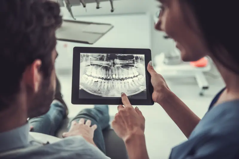
A panoramic image rotates around the head to create a broad view of both jaws, teeth and nearby structures. It can support assessment of tooth development, unerupted teeth, general jaw anatomy and selected pathology.
Its broad coverage comes with less fine detail than intraoral images. A panoramic image may show that a region needs investigation but not replace bitewings for early cavities or a periapical image for a specific root.
Patient positioning matters. Movement, jewellery and posture can create distortion or ghost images.
When is three-dimensional imaging considered?
Cone-beam computed tomography creates a three-dimensional data set. It can be valuable for selected complex root canals, impacted teeth, surgical planning, trauma or pathology.
It generally involves more exposure than a small intraoral image and should not be used as routine screening. The field of view and resolution should be limited to the clinical question.
The prescribing dentist must decide whether the additional information justifies the exposure and whether the scan requires interpretation by someone with relevant expertise.
How often are dental X-rays needed?
No universal six-month, annual or multi-year schedule fits everyone. The dentist considers:
- Current symptoms and clinical findings
- Previous and active cavities
- Periodontal disease and bone monitoring
- Age and tooth development
- Existing restorations
- Dry mouth, diet and fluoride exposure
- Trauma or planned treatment
- Availability and quality of previous images
Routine screening without clinical examination is not appropriate. A risk-based interval can become longer or shorter as circumstances change.
Why might a new dentist request old images?
Previous images can answer current questions and provide a comparison for bone levels, lesions and restoration changes. Health Canada advises attempting to obtain existing information when a patient changes dentists rather than automatically repeating it.
Not every old image remains diagnostically useful. It may not show the required region, may be outdated for an active concern or may have poor quality. The new dentist should explain why another image is necessary.
New symptoms or a changed clinical finding may justify a different view even after records have been transferred. The decision should connect the new exposure to a specific diagnostic question.
Patients can authorize transfer of dental records according to office and regulatory processes.
How much radiation is involved?
Dose depends on image type, equipment, settings, field size and patient anatomy. Health Canada states that the risk from each dental exposure is generally low and that properly conducted dental imaging has a low dose compared with many other medical imaging procedures.
Low risk is not zero risk. Radiation-protection principles require justification, optimized technique, appropriate receptors, collimation and quality assurance to avoid repeats.
Comparisons with flights or background radiation can provide context but vary by location and assumptions. Ask which image is proposed and what it will change instead of relying on a single generic number.
What does ALARA mean?
ALARA means keeping radiation “as low as reasonably achievable” while obtaining necessary diagnostic information. An image with too little exposure to be diagnostic may need repeating and does not serve the patient.
Optimization includes choosing the smallest useful field, appropriate settings, correct positioning and maintained equipment. It also includes not ordering an image when examination and existing records already answer the question.
The principle balances information and exposure rather than pursuing zero dose at the expense of diagnosis.
What about pregnancy?
Tell the dental team if you are pregnant or may be pregnant. Urgent infection and pain should not be ignored. Health Canada notes that fetal exposure from dental X-rays is extremely low because the abdomen is not directly exposed, but the dentist still weighs benefit and risk and uses appropriate precautions.
Elective images without current benefit may be deferred, while a necessary image for urgent diagnosis may be appropriate. The decision depends on the clinical question.
Do not assume that pregnancy requires delaying all dental assessment or treatment.
Why are children imaged selectively?
Children’s mouths change quickly, and images can show cavities between touching teeth, permanent tooth development and trauma. At the same time, children deserve particular attention to field size, exposure settings and thyroid protection when it does not interfere with the required image.
Images are selected from risk and findings rather than age alone. A child with open contacts and low cavity risk may need fewer bitewings than one with previous interproximal decay.
Parents can ask what the image will show and how the result could alter prevention or treatment.
Can I refuse an X-ray?
Patients can make informed decisions, including refusal. The dentist should explain purpose, benefit, risk, alternatives and the limitation created by proceeding without the image.
Sometimes care can be modified or monitored. In other situations, the dentist cannot diagnose or provide a procedure safely without necessary information. Refusal does not obligate a clinician to perform treatment below the standard of care.
Ask whether old images or a different modality could answer the question.
Imaging should answer a question
A useful recommendation sounds specific: “This bitewing will assess a contact that cannot be seen,” or “This periapical image will evaluate the root and surrounding bone.” Generic statements that everyone gets the same set do not explain clinical need.
To learn about Regal Heights Dental Centre’s approach, review our digital dental X-rays and imaging service or contact our team with questions about previous records or a recommended image.
Frequently asked questions
How often should dental X-rays be taken?
There is no single schedule for everyone. Symptoms, age, cavity and gum risk, previous findings and treatment needs determine timing.
Are dental X-rays safe during pregnancy?
Tell the dentist if you are or may be pregnant. Health Canada notes that fetal exposure from dental imaging is extremely low when the abdomen is not directly exposed, but each image still needs clinical justification and appropriate technique.
Why can’t the dentist see a cavity without an X-ray?
Contact areas between back teeth and structures below the gum or inside bone are not fully visible during examination. Bitewing or periapical images may reveal hidden disease.
Does digital imaging have no radiation?
Digital dental radiography still uses ionizing radiation. Modern receptors and optimized equipment can reduce the dose required, but exposure is not zero.